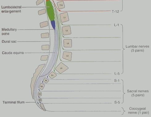
Diagram: The Cauda Equina.
At the level of the thirteenth rib, the human spinal chord terminates in a bulge (the lumbosacral enlargement).
All remaining spinal nerves emerge from the lumbosacral enlargement. These remaining spinal nerves
have the appearance of a horse's tail.
Hence the name. (Cauda equina is Latin for Horse's Tail).
The Clinical significance of the Cauda equina is that
lumbar herniated discs (though exceedingly painful and capable of causing a degree of loss of motor control
to the bladder, bowel, and leg) are unlikely to cause total paraplegia -it takes a spinal fracture to do that.
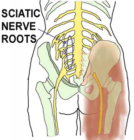
Diagram: The Siatic Nerve and its Roots
The Siatic Nerve originates from five spinal nerve roots: Lumbar nerves L4, L5, and Sacral Nerves S1, S2, and S3.
It is rare for siatic nerve roots other than L4 and L5 to be damaged by lumbar disc herniation.
Nerve Root L4 can be squeezed when the disc between lumbar vertebrae L4 and L5 herniates.
The symptoms of damage to nerve L4 are:-
Difficulty in lifting the big toe off the ground.
Loss of sensation and/or "pins and needles" in the big toe.
Pain in the back of the thigh and the side of the calf.
Nerve Root L5 can be squeezed when the disc between lumbar vertebra L5
and the first sacral vertebra herniates.
The symptoms of damage to nerve L5 are:-
Weakness when trying to put full weight on the ball of the toe and
lift the heal off the ground.
An increased Achilles tendon jerk.
Loss of sensation and/or "pins and needles" in the side of the foot and heel.